
中文摘要:
尽管我们对斑马鱼心脏再生的理解取得了许多进展,但研究较少的一个方面是再生心肌细胞如何侵入和替代含有胶原蛋白的受伤组织。在这里,我们对心肌细胞侵袭的过程进行了深入分析。我们观察到突出的边界区心肌细胞和巨噬细胞之间的密切相互作用,并表明巨噬细胞对于伤口边界区的细胞外基质重塑和心肌细胞突出到受伤区域至关重要。单细胞 RNA 测序揭示了编码膜锚定基质金属蛋白酶的 mmp14b 在边界区的几种细胞类型中的表达。遗传性 mmp14b 突变导致巨噬细胞募集减少、胶原蛋白降解,随后心肌细胞突出到受伤组织中。此外,心肌细胞特异性过表达 mmp14b 足以增强心肌细胞侵袭受伤组织和沿伤口顶端表面的渗透。总而言之,我们的数据为心脏再生过程中心肌细胞侵袭含胶原的受伤组织的机制提供了重要的见解。
英文摘要:
Despite numerous advances in our understanding of zebrafish cardiac regeneration, an aspect that remains less studied is how regenerating cardiomyocytes invade and replace the collagen-containing injured tissue. Here, we provide an in-depth analysis of the process of cardiomyocyte invasion. We observe close interactions between protruding border-zone cardiomyocytes and macrophages, and show that macrophages are essential for extracellular matrix remodeling at the wound border zone and cardiomyocyte protrusion into the injured area. Single-cell RNA-sequencing reveals the expression of mmp14b, encoding a membrane-anchored matrix metalloproteinase, in several cell types at the border zone. Genetic mmp14b mutation leads to decreased macrophage recruitment, collagen degradation, and subsequent cardiomyocyte protrusion into injured tissue. Furthermore, cardiomyocyte-specific overexpression of mmp14b is sufficient to enhance cardiomyocyte invasion into the injured tissue and along the apical surface of the wound. Altogether, our data provide important insights into the mechanisms underlying cardiomyocyte invasion of the collagen-containing injured tissue during cardiac regeneration.
论文信息:
论文题目:Border-zone cardiomyocytes and macrophages regulate extracellular matrix remodeling to promote cardiomyocyte protrusion during cardiac regeneration
期刊名称:Nature Communications
时间期卷:16, Article number: 3823 (2025)
在线时间:2025年4月23日
DOI:doi.org/10.1038/s41467-025-59169-4
产品信息:
货号:CP-005-005
规格:5ml+5ml
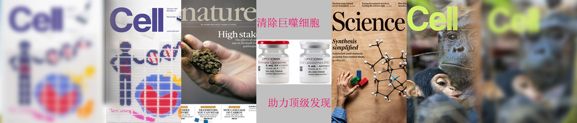
品牌:Liposoma
产地:荷兰
名称:Clodronate Liposomes and Control Liposomes
办事处:Target Technology(靶点科技)
氯膦酸盐脂质体斑马鱼巨噬细胞。斑马鱼心脏冷冻损伤后,多种细胞类型协调对损伤的反应。例如,内皮细胞、心内膜细胞和心外膜细胞已被证明可提供生长因子和信号分子,以促进受伤组织的血运重建和心肌细胞再生。成纤维细胞主要起源于心外膜,已被证明在心脏再生过程中沉积 ECM 以支持心脏,并且对损伤后心肌细胞增殖至关重要。近年来,免疫细胞在促进心脏再生过程中的重要性也得到了认可。特别是,巨噬细胞已被证明在斑马鱼、蝾螈和新生小鼠心脏的损伤再生中起着重要作用。巨噬细胞已被证明直接促进和调节冷冻损伤心脏中瘢痕/ECM 的组成。此外,氯膦酸盐脂质体给药(荷兰Liposoma,清除巨噬细胞)或使用遗传模型消耗巨噬细胞会导致 CM 增殖和再生心脏新生血管形成缺陷。虽然很明显,许多(如果不是全部)心肌细胞类型在心脏再生过程中起着至关重要的作用,但它们对冷冻损伤后心肌细胞纤维化组织重新填充的贡献在很大程度上是未知的。氯膦酸盐二钠脂质体清除巨噬细胞在斑马鱼心脏冻伤模型巨噬细胞功能研究,荷兰Liposoma巨噬细胞清除剂Clodronate Liposomes见刊于Nature Communications:斑马鱼交界区心肌细胞和巨噬细胞调节细胞外基质重塑,促进心脏再生过程中心肌细胞突出。

Liposoma巨噬细胞清除剂Clodronate Liposomes氯膦酸二钠脂质体的材料和方法:

Ventricles from Tg(mpeg:NTR-YFP) adult fish were cryoinjured as described above. Cryoinjured fish were then incubated in a 5 μm nifurpirinol or DMSO-control water bath from 4 to 6 days following cryoinjury (replenished daily). Hearts were extracted at 7 dpci and subjected to immunostaining with GFP antibody to check macrophage ablation efficiency and phalloidin staining to quantify CM protrusion at the border zone as described above.
To ablate resident macrophages, Tg(mpeg1:EGFP) adult fish were IP injected with 10 μl clodronate liposomes (5 mg/ml) (Liposoma, Amsterdam, The Netherlands) or PBS 8 days prior to cryoinjury. Hearts were extracted at 7 or 10 dpci followed by cryosection, CHP staining, and phalloidin staining, as described above. Quantification of CHP intensity and CM protrusion at the border zone were performed as described above.
To ablate macrophages at later timepoints, Tg(mpeg1:EGFP) adult fish were IP injected with 10 μl clodronate liposomes (5 mg/ml) (Liposoma, Amsterdam, The Netherlands) or PBS control liposomes at 3, 6, and 9 dpci before collection of hearts at 10 dpci. To study the impact of macrophage ablation on mmp14b overexpression in CMs, Tg(hsp70l:loxP-EGFP-loxP-mmp14b-P2A-tagBFP); Tg(−5.1myl7:CreERT2) adult fish were IP injected with clodronate liposomes or PBS liposomes 1 day prior to cryoinjury and every 4 days afterwards. Tamoxifen/ethanol injections and heat shock were performed to induce mmp14b overexpression as described above. Hearts were extracted at 10 or 21 dpci followed by cryosection and phalloidin or MHC staining as described above. Quantification of CM protrusion at the border zone and CM cortical coverage were performed as described above.