中文摘要:
目前纳米颗粒(NPs)已被研发应用于药物递送与分子成像领域。然而,纳米颗粒往往在抵达靶标前就被机体截留,导致靶向效率和信噪比偏低,且易在肺、肝脏、肾脏、脾脏等器官中蓄积。
目前的解决办法是反复优化纳米颗粒的表面特性与给药策略,但该流程耗时冗长,还需要在不同时间点进行脏器解剖。为解决这一痛点,本研究提出一种快速迭代研究方案:利用活体动物 X 射线荧光(XRF)成像,系统性评估纳米颗粒的体内分布。
我们将该方法应用于钼基纳米颗粒与荷兰Liposoma氯膦酸盐脂质体,通过瞬时耗竭巨噬细胞实现肿瘤靶向,有效减少了纳米颗粒在肺部和肝脏的蓄积,最终实现肿瘤病灶检出。X 射线荧光计算机断层扫描(XFCT)可提供纳米颗粒在肿瘤内部分布的三维空间信息。
研究通过多尺度成像技术(掺杂荧光染料纳米颗粒)及基因表达分析开展纳米毒理学特征研究,对上述结果进行了验证。XRF 成像技术有望推动临床前药代动力学研究中治疗与诊断手段的发展。
英文摘要:
Nanoparticles (NPs) are currently developed for drug delivery and molecular imaging. However, they often get intercepted before reaching their target, leading to low targeting efficacy and signal-to-noise ratio. They tend to accumulate in organs like lungs, liver, kidneys, and spleen. The remedy is to iteratively engineer NP surface properties and administration strategies, presently a time-consuming process that includes organ dissection at different time points. To improve this, we propose a rapid iterative approach using whole-animal x-ray fluorescence (XRF) imaging to systematically evaluate NP distribution in vivo. We applied this method to molybdenum-based NPs and clodronate liposomes for tumor targeting with transient macrophage depletion, leading to reduced accumulations in lungs and liver and eventual tumor detection. XRF computed tomography (XFCT) provided 3D insight into NP distribution within the tumor. We validated the results using a multiscale imaging approach with dye-doped NPs and gene expression analysis for nanotoxicological profiling. XRF imaging holds potential for advancing therapeutics and diagnostics in preclinical pharmacokinetic studies.
论文信息:
论文题目:Iterative nanoparticle bioengineering enabled by x-ray fluorescence imaging
期刊名称:Science Advances
时间期卷:Vol 10, Issue 12(2024)
在线时间:2024年3月22日
DOI: 10.1126/sciadv.adl2267
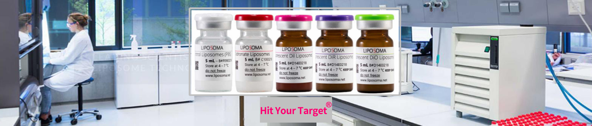
产品信息:
货号:CP-005-005
规格:5ml+5ml
品牌:Liposoma
产地:荷兰
名称:Clodronate Liposomes&Control Liposomes
办事处:靶点科技
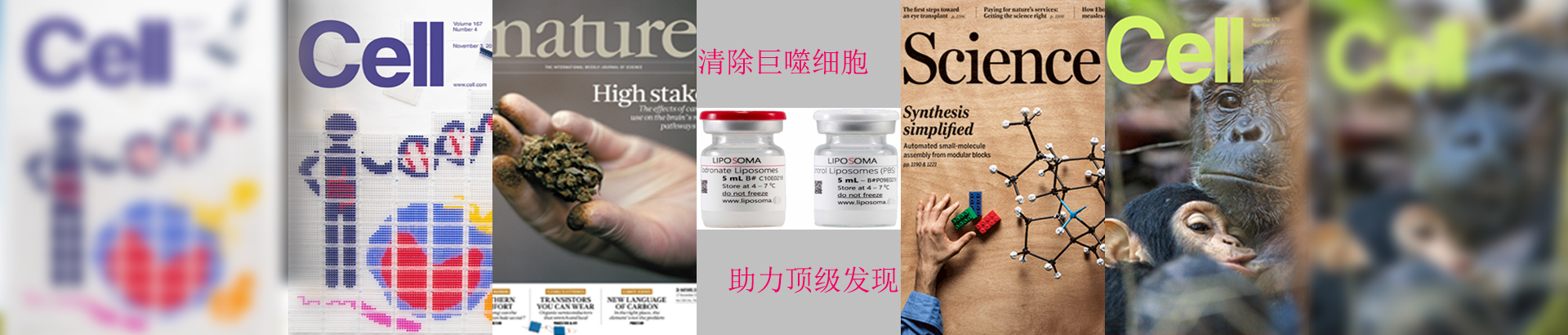
Clodronate Liposomes氯膦酸盐脂质体在肿瘤模型种清除肝脏和脾脏巨噬细胞,实现纳米颗粒(NPs)靶向肿瘤细胞递送。荷兰Liposoma巨噬细胞清除剂ClodronateLiposomes见刊于Science Advances:基于 X 射线荧光成像的纳米颗粒迭代生物工程改造。

Liposoma巨噬细胞清除剂Clodronate Liposomes氯膦酸二钠脂质体清除巨噬细胞的材料和方法:
Ammonium heptamolybdate [AHM; (NH4)6Mo7O24·4H2O], PVP (55 kDa), Cy5.5 mono NHS ester (Cy5.5-NHS), (3-aminopropyl) triethoxysilane (APTES; C9H23NO3Si, 99%), (TEA; C6H15N, ≥99%), dimethyl sulfoxide (DMSO; C2H6OS·H2O, ≥99%), tetraethyl orthosilicate (TEOS; C8H20O4Si ≥ 99%), and EA (C2H7NO, ≥99%) were all purchased from Sigma-Aldrich (Stockholm, Sweden). Ethanol (EtOH) absolute (≥99.8%) was obtained from VWR (Stockholm, Sweden). A MilliQ reference water purification system (Merck Millipore) was used for deionized (DI) water. Clodronate-encapsulated liposomes and empty (control) liposomes were purchased from Liposoma BV (Amsterdam, The Netherlands).
巨噬细胞清除材料和方法文献截图:
