中文摘要:
α- 突触核蛋白(α-syn)原纤维会在帕金森病患者体内蓄积,并在细胞间传播,通过模板诱导蛋白错误折叠,进而诱发神经退行性病变。α- 突触核蛋白原纤维进入健康神经元是该病理过程的关键环节。
本研究全面筛选了可与 α- 突触核蛋白原纤维结合的膜蛋白质组,鉴定出mGluR4与NPDC1两种黑质表面蛋白,二者能够结合并介导 α- 突触核蛋白原纤维的细胞内吞。
向野生型小鼠纹状体内注射 α- 突触核蛋白原纤维,会造成黑质多巴胺能神经元损伤;而敲除Grm4或Npdc1任一基因,均可对多巴胺能神经元起到保护作用。研究发现,mGluR4 与 NPDC1 可形成复合物,并调控 mGluR4 的生理功能。
同时缺失Grm4和Npdc1的培养神经元,无法结合 α- 突触核蛋白原纤维,也不会出现磷酸化 α- 突触核蛋白蓄积及突触丢失现象。Grm4、Npdc1 双杂合转基因小鼠的黑质神经元可免受 α- 突触核蛋白原纤维的损伤,证实两个基因之间存在遗传互作。
在 α- 突触核蛋白 A53T 转基因模型小鼠中,Grm4与Npdc1双杂合状态可显著延长小鼠生存期、改善运动功能,并维持脊髓运动神经元数量。
综上,细胞表面 mGluR4–NPDC1 复合物 参与介导了 α- 突触核蛋白驱动的神经退行性病变进程。
英文摘要:
α-Syn fibril entry into healthy neurons is a key step. Here, we comprehensively assessed the membrane proteome for α-syn fibril binding. We identified mGluR4 and NPDC1 as nigral surface proteins binding and internalizing α-syn fibrils. While striatal α-syn fibril injection led to nigral dopamine neuron loss in wild type mice, deletion of either Grm4 or Npdc1 provided protection of dopamine neurons. We observed mGluR4 and Npdc1 to form a complex regulating mGluR4 function. Cultured neurons lacking both Grm4 and Npdc1 fail to bind α-syn fibrils, to accumulate phosphorylated α-syn and to lose synapses. Transheterozygous Grm4, Npdc1 mice showed protection of nigral neurons from α-syn fibrils, demonstrating genetic interaction. For transgenic α-syn A53T mice, double Grm4, Npdc1 heterozygosity increased mouse survival, motor function and spinal motoneuron number. Thus, a cell surface mGluR4–NPDC1 complex participates in α-syn neurodegeneration.
论文信息:
论文题目:mGluR4–NPDC1 complex mediates α-synuclein fibril-induced neurodegeneration
期刊名称:Nature Communications
时间期卷:17, Article number: 994(2026)
在线时间:2025年12月25日
DOI: doi.org/10.1038/s41467-025-67731-3
产品信息:
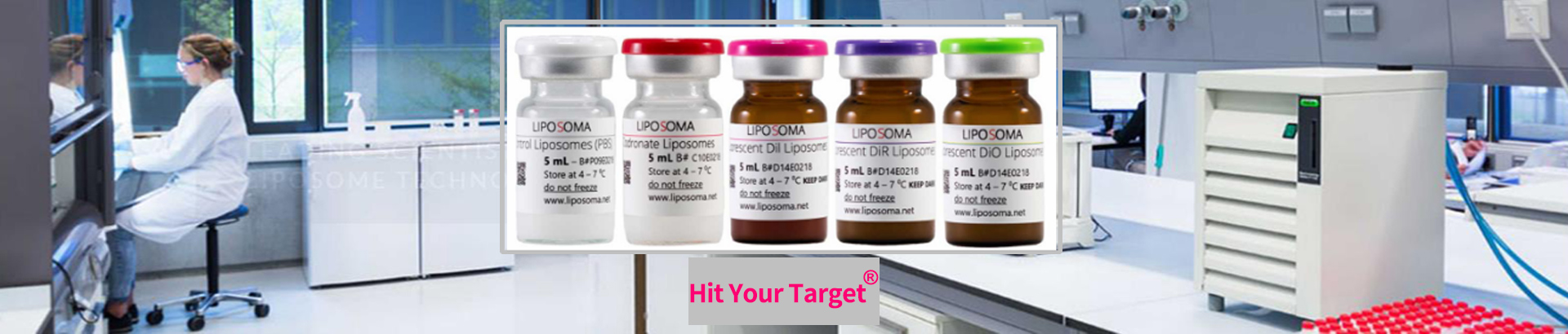
货号:HECA500
规格:500ml
品牌:Brainbits
产地:美国
名称:BrainBits 无钙培养液 E 型
办事处:靶点科技
Brainbits胚胎型神经培养液见刊于Nature Communications:mGluR4–NPDC1 复合物介导 α- 突触核蛋白原纤维诱导的神经退行性病变

Brain bits胚胎E型培养基的材料和方法:
Cortical cells were isolated from the cortices of E16-17 mouse embryos as reported previously49. The embryos were decapitated, and the anterior cortex was dissected out under a stereotactic microscope using BrainBits Hibernate E minus calcium medium, kept on ice. The tissue was then digested with a freshly prepared enzyme solution (Mg/Ca-free HBSS containing 20 U/ml Papain (Worthington LK003178), 3 mM EDTA (AmericanBio), 2 mM CaCl₂ (VWR E506), and 1 mg/ml DNAse (Sigma DN25)) at 37 °C for 30 min. After digestion, the cell suspension was filtered through a 40 µm cell strainer (Corning, #352340), and the cells were counted and diluted to a concentration of 1 × 10⁵ cells/ml. A 100 µl volume of this suspension was plated onto a PDL-coated 96-well Black/Clear Flat Bottom Plate. The plates were incubated at 37 °C with 5% CO₂, and the medium was half-changed every 7 days.
For the binding assay, PFF was added at DIV 17-18 and incubated at 4 °C or 37 °C for 2 h. For the α-syn phosphorylation and synaptic loss assays, PFF were added at DIV 10 and incubated at 37 °C for 7 days.
材料和方法文献截图:
