肝脏再生模型里面,巨噬细胞如何发挥功能?一个重要的手段就是使用巨噬细胞清除氯膦酸盐脂质体Clodronate Liposomes。金标准产品就是荷兰Liposoma的CP-005-005。可以参考如下文献。
论文信息:
论文题目:NLRP3 inflammasome constrains liver regeneration through impairing MerTK-mediated macrophage efferocytosis
期刊名称:Science Advances
时间期卷:Vol 1, Issue 1(2025)
在线时间:2025年1月1日
DOI:doi.org/10.1126/sciadv.adq5786
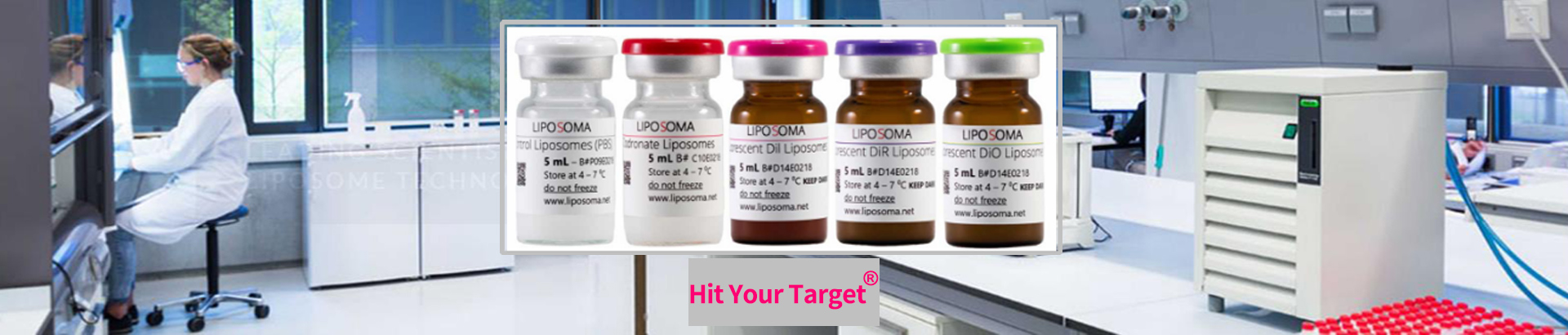
产品信息:
货号:CP-005-005
规格:5ml+5ml
品牌:Liposoma
产地:荷兰
名称:Clodronate Liposomes&Control Liposomes
办事处:靶点科技

肝脏切除前4h,尾静脉注射清除剂C-005和对照试剂P-005。48h后取肝脏做免疫组化IHC,染色F4/80。IHC显示,荷兰Liposoma的CP-005-005巨噬细胞清除套装,有效的清除了肝脏巨噬细胞。
Clodronate Liposomes氯膦酸盐脂质体在小鼠创肝脏再生模型种清除肝脏巨噬细胞。荷兰Liposoma巨噬细胞清除剂ClodronateLiposomes见刊于Science Advances:NLRP3 炎症小体通过抑制 MerTK 介导的巨噬细胞胞葬作用,进而限制肝脏再生。

Liposoma巨噬细胞清除剂Clodronate Liposomes氯膦酸二钠脂质体清除巨噬细胞的材料和方法:
To generate the PHx model, mice were subjected to 70% PHx as our previous description . For macrophage depletion, 200 μl of CLs (Liposoma) and control liposomes were intraperitoneally injected 4 hours before PHx. For hepatic NLRP3 overexpression, the mice were injected with AAV8-NLRP3 (AAV-OE) or AAV-8-NTC (AAV-BL) (General Biology) at a dosage of 1 × 10¹² vector genomes (vg) per mouse intravenously 20 days before PHx operation. For NLRP3 inhibition, MCC950 (TargetMol, no. T3701) was administrated intraperitoneally to a concentration of 20 μg/kg 2 hours before PHx. For MerTK inhibition, UNC2025 (TargetMol, no. T7007) was administrated intraperitoneally to a concentration of 50 mg/kg 2 hours before and 24 hours after PHx. For IL-1β inhibition, IL-1β neutralizing antibody (R&D Systems, no. AF-401-NA) was administrated intraperitoneally to a concentration of 10 mg/kg 4 hours before PHx. For macrophage depletion, CLs (200 μl; Yeasen, no. 40337ES08) and control liposomes (200 μl; Yeasen, no. 40338ES08) were administered by tail vein injection 4 hours before PHx. The mice were humanly euthanized at indicated time after PHx. Blood and liver tissue samples were collected for examination. All animal experiments were approved by the Ethics Committee Medical College of Qingdao University (approval no. QDU-AEC-2021151).
巨噬细胞清除会延缓肝脏再生进程,并抑制肝细胞增殖。


图4.巨噬细胞清除会延缓肝脏再生与肝细胞增殖
(A) 本实验中巨噬细胞清除联合部分肝切除(PHx)体内造模流程示意图。
(B) 对照组(NC)与巨噬细胞清除组(CLs)小鼠在 PHx 术后第 2 天肝脏组织 F4/80 抗原免疫组化(IHC)染色代表性图片(比例尺,100 μm)。
(C) PHx 术后对照组与巨噬细胞清除组小鼠生存曲线(每组 n=10)。
(D) PHx 术后各时间点小鼠肝重 / 体重比(每组 n=4~6)。
(E) 肝脏组织切片中 Ki67 阳性、BrdU 阳性细胞免疫荧光(IF)染色代表性图片(比例尺,100 μm)。
(F、G) 肝脏切片中 Ki67⁺、BrdU⁺细胞数量统计(每组 n=5)。
(H) PHx 术后 48 小时,对照组(NC)与巨噬细胞清除组(CLs)小鼠肝脏 CCND1、PCNA、前体半胱天冬酶 3(Pro-Cas3)、活化剪切型半胱天冬酶 3(C-Cas3)蛋白表达的蛋白质免疫印迹(Western blot)检测结果(每组 n=4)。
(I、J) 末端脱氧核苷酸转移酶介导的 dUTP 缺口末端标记(TUNEL)染色代表性图片及阳性细胞定量统计(比例尺,100 μm)(每组 n=5)。